Sonography
Sonography or ultrasound is used to evaluate the abdomen. High-frequency sound waves are used to create an image that helps to diagnose a medical condition such as an enlarged organ, liver disease, or kidney stone, Jariwala Women's Hospital has equipped with modern facilities that make sonography easy for patients and help doctors to diagnose accurately.
What We Do
Sonography, or ultrasound, is a safe, painless imaging technique that uses sound waves to create real-time images of internal organs and developing babies during pregnancy. We use advanced sonography to monitor fetal growth, assess reproductive organs, diagnose conditions, and guide fertility treatments. Whether it’s tracking your baby’s development or evaluating your health, our expert team ensures accurate imaging and compassionate care at every step.
How We Help You
We provide high-quality sonography services to give you clear answers and peace of mind. Whether you're monitoring your pregnancy, evaluating fertility concerns, or checking reproductive health, our skilled specialists use advanced ultrasound technology for precise and safe imaging. We explain every step of the process, ensure your comfort, and help you understand the results—so you feel confident, cared for, and fully informed throughout your journey.
Procedure
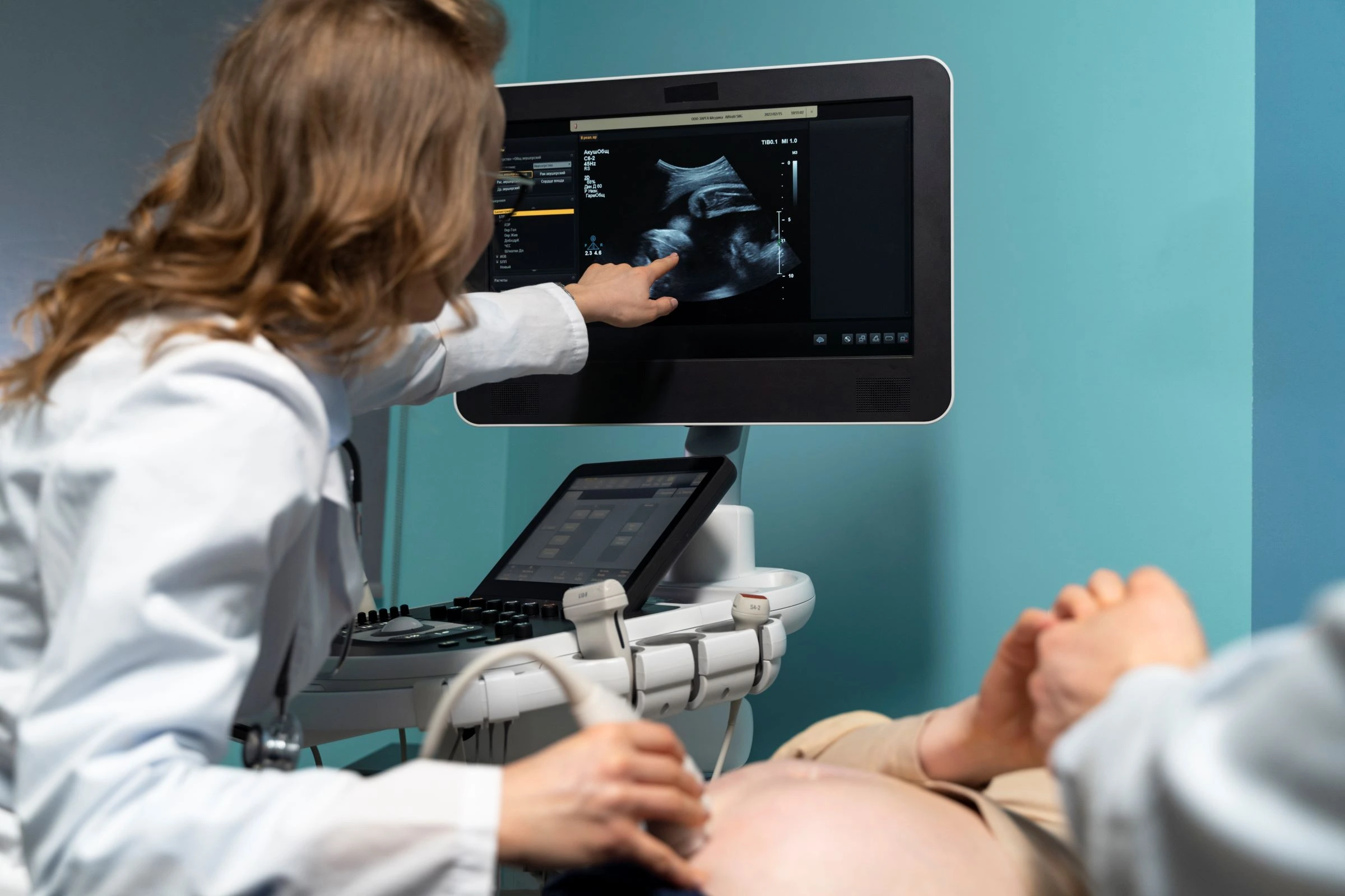
The sonography procedure involves applying a special gel to the skin over the area being examined. A handheld device called a transducer is then moved over the area, sending sound waves into the body and capturing the echoes that bounce back to create real-time images. The process is completely safe, non-invasive, and usually takes 15–30 minutes. It helps in monitoring pregnancy, evaluating internal organs, and guiding fertility or diagnostic care.